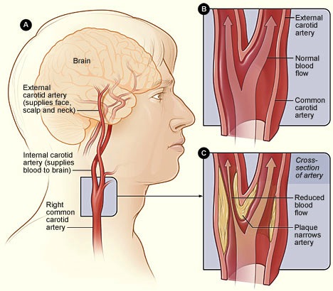
Carotid (kuh-ROT-id) ultrasound is a safe, painless procedure that uses sound waves to examine the structure and function of the carotid arteries in your neck. Your two carotid arteries are located on each side of your neck. Carotid arteries deliver blood from your heart to your brain.
Carotid ultrasound is usually used to test for blocked or narrowed carotid arteries, which can indicate an increased risk of stroke. Results from a carotid ultrasound can help your doctor determine what kind of treatment you may need to lower your risk of stroke.
Why It’s Done
The primary purpose of a carotid ultrasound is to test for narrowed carotid arteries that indicate an increased risk of stroke.
Narrowing of carotid arteries is usually caused by plaque — a buildup of fat, cholesterol, calcium and other substances that circulate in the bloodstream. Early detection of narrowed carotid arteries enables your doctor to begin treatments to improve blood flow to your brain and decrease your risk of stroke.
Your doctor may recommend a carotid ultrasound if you have medical conditions that increase the risk of stroke, including:
• High blood pressure
• Diabetes
• High cholesterol
• Family history of stroke or heart disease
• Recent transient ischemic attack (TIA) or stroke
• Abnormal sound in carotid arteries (bruit), detected by your doctor using a stethoscope
You’ll have a Doppler ultrasound that evaluates the blood flow through your carotid arteries.
A carotid ultrasound may be used in combination with other tests to screen for narrowed or blocked blood vessels in other areas of your body, including:
• Abdominal ultrasound. You may have an abdominal ultrasound to test for conditions affecting the blood vessels or organs in your abdominal area.
• Ankle-brachial index test. This test measures and compares your ankle’s blood pressure and your arm’s blood pressure. The test can indicate reduced or blocked blood flow to your legs.
Other uses of carotid ultrasound
Your doctor also may order a carotid ultrasound to:
• Evaluate the structure and function of the artery after surgery to remove plaques (carotid endarterectomy)
• Evaluate the placement and treatment effect of a stent, a mesh tube used to improve blood flow through an artery by mechanically decreasing the narrowing
• Locate a collection of clotted blood (hematoma) that may inhibit blood flow
• Detect other abnormalities in the structure of a carotid artery that may disrupt blood flow
How You Prepare
You can take the following steps to prepare for your appointment:
• Call the day before the exam to confirm the time and location of the exam.
• Wear a comfortable shirt with no collar or an open collar.
• Don’t wear a necklace or dangling earrings.
Unless your doctor or the radiology lab provides special instructions, you shouldn’t need to make any other preparations.
What you Can Expect
A technician (sonographer) conducts the test with a small, hand-held device called a transducer. The transducer emits sound waves and records the echo as the waves bounce off tissues, organs and blood cells.
A computer translates the echoed sound waves into a live-action image on a monitor. In a Doppler ultrasound, the information about the rate of blood flow is translated into a graph.
A carotid ultrasound usually takes about 30 minutes.
During the procedure
You’ll likely lie on your back during the procedure. The ultrasound technician (sonographer) may gently adjust the position of your head to improve access to the side of your neck.
The sonographer will apply a warm gel to your skin above the site of each carotid artery. The gel helps eliminate the formation of air pockets between your skin and the transducer. The sonographer then gently presses the transducer against the side of your neck in order for the instrument to send and receive sound waves.
You shouldn’t feel any discomfort during the procedure. If you do, tell the sonographer.
Cardiac Specialty Care
• Structural Heart Disease
• TAVR
• CardioMEMS (Heart Failure)
• PFO Closure
• TAVR
• CardioMEMS (Heart Failure)
• PFO Closure
• Coronary Intervention
• Complex Higher-Risk (And Indicated) Patients (CHIP) Angioplasty
• Atherectomy
• Impella and ECMO Support
• Complex Higher-Risk (And Indicated) Patients (CHIP) Angioplasty
• Atherectomy
• Impella and ECMO Support
• Peripheral Angioplasty
• Varicose Vein Treatment (Venous Ablation)
• DVT thrombectomy - IVC filter
• Carotid Stenting
• Varicose Vein Treatment (Venous Ablation)
• DVT thrombectomy - IVC filter
• Carotid Stenting
• Rhythm Management
• Pacemaker
• Holter Monitoring
• Exercise Stress Test
• Echocardiography
• Nuclear Stress Test
• Enhanced External Counterpulsation (EECP)
• Pacemaker
• Holter Monitoring
• Exercise Stress Test
• Echocardiography
• Nuclear Stress Test
• Enhanced External Counterpulsation (EECP)
