Stent
Astent is a tiny tube placed into an artery, blood vessel, or other hollow structure (such as one that carries urine) to hold it open.
Description
When a stent is placed into the body, the procedure is called stenting. There are different kinds of stents. Most are made of a metal or plastic mesh-like material. However, stent grafts are made of fabric. They are used in larger arteries.
• An intraluminal coronary artery stent is a small metal mesh tube that expands in the artery. It is placed inside a coronary artery after balloon angioplasty to prevent the artery closing again.
• A drug-eluting stent is a stent that is coated with a medicine that helps prevent the arteries from re-closing. Like other coronary artery stents, it is left in place in the artery.
Doctors place stents in arteries as part of a procedure called percutaneous coronary intervention (PCI), sometimes referred to as coronary angioplasty.
To place a stent, your doctor will make a small opening in a blood vessel in your groin (upper thigh), arm, or neck.Through this opening, your doctor will thread a thin, flexible tube called a catheter. The catheter will have a deflated balloon at its tip.A stent is placed around the deflated balloon. Your doctor will move the tip of the catheter to the narrow section of the artery or to the aneurysm or aortic tear site.
Special x-ray movies will be taken of the tube as it’s threaded through your blood vessel. These movies will help your doctor position the catheter.
For Arteries Narrowed by Plaque
Your doctor will use special dye to help show narrow or blocked areas in the artery. He or she will then move the catheter to the area and inflate the balloon.
As the balloon inflates, it pushes the plaque against the artery wall. This widens the artery and helps restore blood flow. The fully extended balloon also expands the stent, pushing it into place in the artery.
The balloon is deflated and pulled out along with the catheter. The stent remains in your artery. Over time, cells in your artery grow to cover the mesh of the stent. They create an inner layer that looks like the inside of a normal blood vessel.
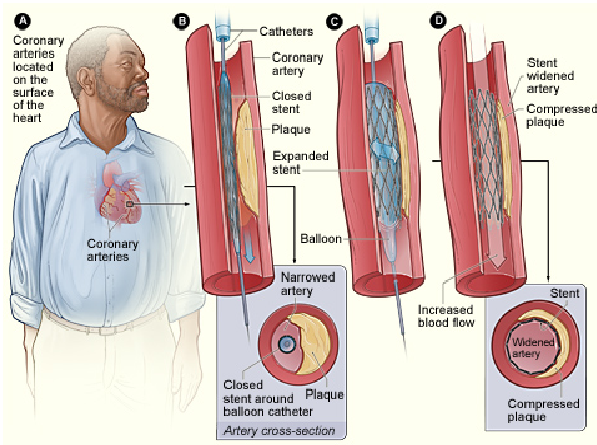
Coronary Artery Stent Placement
Figure A shows the location of the heart and coronary arteries. Figure B shows the deflated balloon catheter and closed stent inserted into the narrow coronary artery. The inset image shows a cross-section of the artery with the inserted balloon catheter and closed

stent. In figure C, the balloon is inflated, expanding the stent and compressing the plaque against the artery wall. Figure D shows the stent-widened artery. The inset image shows a cross-section of the compressed plaque and stent-widened artery.
A very narrow artery, or one that’s hard to reach with a catheter, may require more steps to place a stent. At first, your doctor may use a small balloon to expand the artery. He or she then removes the balloon.
The small balloon is replaced with a larger balloon that has a collapsed stent around it. At this point, your doctor can follow the standard process of compressing the plaque and placing the stent.
Doctors use a special filter device when doing PCI and stent placement on the carotid arteries. The filter helps keep blood clots and loose pieces of plaque from traveling to the brain during the procedure.
For Aortic Aneurysms
The procedure to place a stent in an artery with an aneurysm is very similar to the one described above. However, the stent used to treat an aneurysm is different. It’s made out of pleated fabric instead of metal mesh, and it often has one or more tiny hooks.
The stent is expanded to fit tight against the artery wall. The hooks latch on to the wall of the artery, holding the stent in place.
The stent creates a new inner lining for that portion of the artery. Over time, cells in the artery grow to cover the fabric. They create an inner layer that looks like the inside of a normal blood vessel.
Cardiac Specialty Care
• Structural Heart Disease
• TAVR
• CardioMEMS (Heart Failure)
• PFO Closure
• TAVR
• CardioMEMS (Heart Failure)
• PFO Closure
• Coronary Intervention
• Complex Higher-Risk (And Indicated) Patients (CHIP) Angioplasty
• Atherectomy
• Impella and ECMO Support
• Complex Higher-Risk (And Indicated) Patients (CHIP) Angioplasty
• Atherectomy
• Impella and ECMO Support
• Peripheral Angioplasty
• Varicose Vein Treatment (Venous Ablation)
• DVT thrombectomy - IVC filter
• Carotid Stenting
• Varicose Vein Treatment (Venous Ablation)
• DVT thrombectomy - IVC filter
• Carotid Stenting
• Rhythm Management
• Pacemaker
• Holter Monitoring
• Exercise Stress Test
• Echocardiography
• Nuclear Stress Test
• Enhanced External Counterpulsation (EECP)
• Pacemaker
• Holter Monitoring
• Exercise Stress Test
• Echocardiography
• Nuclear Stress Test
• Enhanced External Counterpulsation (EECP)
